Research Article | | Peer-Reviewed

Determination of the Acidity Constants of Serotonin in the Ground and Excited States Using Spectroscopic Methods

Received: 15 March 2026     Accepted: 24 March 2026     Published: 14 April 2026
Views:       Downloads:
Abstract

Serotonin (5-hydroxytryptamine, 5-HT) is a biologically important neurotransmitter whose photophysical behavior depends on its protonation state. This study investigates the acid-base properties of serotonin in aqueous solution using UV-visible absorption and fluorescence spectroscopies. The ground-state acidity constant (pKa(S0)) was determined spectrophotometrically, while the excited-state acidity constant (pKa*(S1)) was estimated using the Förster thermodynamic cycle. Absorption spectra revealed an isosbestic point, indicating a simple two-state equilibrium between the protonated (R-OH) and deprotonated (R-O⁻) forms. The results show that serotonin behaves as a weak acid in the ground state (pKa(S0) ≈ 10.5–10.6), whereas its acidity increases significantly in the excited state (pKa*(S1) ≈ 4.7) due to electronic redistribution within the indole chromophore. Excited-State Proton Transfer (ESPT) occurs efficiently, influencing both fluorescence intensity and emission wavelength. These findings provide a comprehensive understanding of serotonin’s photophysical behavior and support its use as an intrinsic fluorescent probe for monitoring local pH variations in aqueous or cellular environments. The combination of UV-Vis and fluorescence measurements, with triplicate statistical validation, ensures reproducibility and accuracy of the determined acidity constants. This work contributes to a better understanding of neurotransmitter acid-base behavior under physiologically relevant conditions and demonstrates the potential application of serotonin in fluorescence-based pH sensing and molecular studies.

Published in American Journal of Chemical Engineering (Volume 14, Issue 2)
DOI 10.11648/j.ajche.20261402.11
Page(s) 19-28
Creative Commons

This is an Open Access article, distributed under the terms of the Creative Commons Attribution 4.0 International License (http://creativecommons.org/licenses/by/4.0/), which permits unrestricted use, distribution and reproduction in any medium or format, provided the original work is properly cited.

Copyright

Copyright © The Author(s), 2026. Published by Science Publishing Group

Keywords

Serotonin, UV-Vis Absorption Spectroscopy, Fluorescence Spectroscopy, Acid-Base Equilibrium, pKa, Excited-State Proton Transfer (ESPT), Isosbestic Point

1. Introduction
Serotonin (5-hydroxytryptamine, 5-HT) is a monoamine neurotransmitter widely distributed in both the central and peripheral nervous systems. It plays a central role in regulating physiological processes such as mood, appetite, sleep, pain perception, and gastrointestinal motility . Beyond its neurotransmitter function, serotonin is involved in neuroendocrine signaling, cardiovascular regulation, and immune modulation .
Structurally, serotonin is an indoleamine consisting of an indole ring fused to an ethylamine side chain with a hydroxyl group at the 5-position. This indole motif endows the molecule with characteristic photophysical properties, enabling absorption of ultraviolet (UV) light and emission of fluorescence in aqueous environments . Similar to tryptophan and other indole derivatives, serotonin exhibits π→π* electronic transitions in the UV region, making it naturally fluorescent and a versatile probe for studying local chemical environments .
Fluorescence properties of serotonin are highly sensitive to factors such as solvent polarity, ionic strength, viscosity, and pH. This sensitivity allows monitoring of protonation states and intermolecular interactions in both biological and chemical systems . Fluorescence spectroscopy has been successfully used to study serotonin interactions with proteins, membranes, and nucleic acids, as well as its dynamic behavior in living cells .
A critical factor influencing fluorescence is serotonin’s protonation state, which depends on solution pH. The molecule contains a phenolic hydroxyl group and a primary amine, each capable of undergoing protonation-deprotonation equilibria . Changes in protonation alter electron distribution in the indole chromophore, leading to measurable shifts in absorption and emission spectra. Monitoring these shifts allows determination of the ground-state acidity constant (pKa(S0)) and the excited-state acidity constant (pK*(S1)) .
Upon excitation (S0 → S1), electron density is redistributed within the indole system. This rearrangement typically increases the acidity of the phenolic hydroxyl group, favoring deprotonation in the excited state. Such behavior underlies Excited-State Proton Transfer (ESPT) processes, which can strongly influence fluorescence intensity, spectral position, and interactions with solvent molecules . The Förster thermodynamic cycle provides a framework for linking pKa(S0) and pK*(S1), allowing estimation of excited-state acidity from ground-state measurements and electronic transition energies .
In this study, we focus on the phenolic hydroxyl group as the main acidic site, while the primary amine remains largely protonated over the pH range investigated. UV-visible absorption spectra of serotonin show a prominent band around 278 nm, corresponding to π→π* transitions of the indole chromophore. This wavelength was chosen for both ground- and excited-state pKa determinations and fluorescence measurements because it selectively probes the phenolic group without interference from the amino group .
The present work aims to provide a comprehensive spectroscopic analysis of serotonin in aqueous solution. Using UV-Vis and fluorescence spectroscopies, we determine its acidity constants in both ground and excited states, investigate ESPT processes, and discuss the photophysical mechanisms that govern fluorescence behavior. These insights are critical for understanding serotonin’s interactions at the molecular level and for its potential use as an intrinsic fluorescent probe, particularly for monitoring local pH changes in aqueous or cellular environments . Although ground- and excited-state pKa values of serotonin have been reported, this study provides a refined spectroscopic determination combining UV-Vis and fluorescence measurements with triplicate statistical validation, and explores the protonation behavior under physiologically relevant conditions, offering improved accuracy and reproducibility for potential use in pH-sensing applications.
2. Materials and Methods
2.1. Chemicals and Reagents
Serotonin hydrochloride (≥ 99% purity) was purchased from Alfa Aesar and used without further purification. The compound corresponds to the hydrochloride salt of 5-hydroxytryptamine, with molecular formula C10H12N2O·HCl and molar mass 198.5 g mol-1. Stock solutions were prepared at 1.0 × 10⁻2 M in distilled water and stored in the dark. Working solutions in the micromolar range (10⁻⁵-6×10⁻⁵ M) were prepared by serial dilution immediately before use. All solutions were protected from light and used within 48 hours. Buffer solutions covering the pH range from 2 to 13 were prepared by using 0.05 M phosphate buffer (NaH2PO4 / Na2HPO4) for pH 6-8, 0.05 M acetate buffer (CH3COONa / CH3COOH) for pH 3-5, 0.05 M borate buffer (H3BO3 / Na2B4O7) for pH 9-10.5, and dilute HCl or NaOH solutions for the extreme pH values below 2 or above 13. The pH of all solutions was measured at 25 °C using a calibrated pH meter (±0.01 unit accuracy).
2.2. UV-visible Absorption Spectroscopy
Absorption spectra were recorded on a Cary 100 UV-visible spectrophotometer using quartz cuvettes of 1 cm path length. Measurements were performed in the 200-400 nm wavelength range. Baseline correction was done using the corresponding buffer. Linearity of the Beer-Lambert law was verified for all experimental concentrations to confirm the absence of aggregation or excimer formation. Molar extinction coefficients (ε) were calculated from linear regression of absorbance versus concentration.
2.3. Fluorescence Spectroscopy
Fluorescence measurements were carried out on a PerkinElmer LS 55 spectrofluorometer equipped with 1 cm quartz cuvettes. Excitation and emission slit widths were optimized to minimize inner-filter effects. No correction for inner-filter absorption was applied, as absorbance at λex = 278 nm remained below 0.05 for all samples. Measurements were performed at 25 °C. Emission spectra were recorded from 300 to 450 nm. Data processing, curve fitting, and statistical analysis were performed using OriginPro software. The absorbance at λex = 278 nm remained below 0.05 for all samples. According to Lakowicz (2006), this level is sufficiently low to neglect inner-filter effects, ensuring linearity between concentration and fluorescence intensity.
2.4. Determination of Ground- and Excited-State Acidity Constants
The acidity constants were determined using a combination of spectrophotometric methods. Ground-state pKa(S0) was obtained from the absorbance ratio of protonated (R-OH) and deprotonated (R-O⁻) species at λ = 278 nm, using both the percentage method and the Henderson-Hasselbalch equation:
pH= pKaS0+log [R-O [R-OH] 
The excited-state pKa*(S1) was estimated using the Förster thermodynamic cycle, linking ground-state acidity and electronic transition energies of the protonated and deprotonated forms.
2.5. Experimental Reproducibility
All measurements were performed in triplicate, and standard deviations were calculated. Consistency between UV-Vis and fluorescence determinations ensured reliable analysis of serotonin’s acid-base behavior in both ground and excited states.
3. Results and Discussion
3.1. UV-visible Absorption Spectrum of Serotonin
The UV-visible absorption spectrum of serotonin (5-hydroxytryptamine, 5-HT) in aqueous solution exhibits distinct absorption bands at 200, 219, 275, and 297 nm, corresponding to the π → π* electronic transitions of the indole chromophore (Figure 1) . The most intense absorption at 200 nm (ε = 16,149 M-1 cm-1) reflects a high-energy transition localized on the indole ring, consistent with other indole derivatives, while the band at 219 nm (ε = 14,091 M-1 cm-1) involves additional contributions from the conjugated amine system, indicating extended delocalization within the chromophore . Longer-wavelength transitions at 275 nm (ε = 3,606 M-1 cm-1) and 297 nm (ε = 2,894 M-1 cm-1) are attributed to the phenolic OH group interacting with the indole π system, which is particularly relevant for fluorescence excitation in subsequent pKa and ESPT studies. Excitation in this region efficiently populates the S1 excited state, leading to emission around 335-340 nm . Linearity of absorbance with concentration was confirmed using Beer-Lambert plots, yielding correlation coefficients r2 = 0.999 for all bands. This demonstrates the absence of aggregation or excimer formation in the tested micromolar range (10⁻⁵-6×10⁻⁵ M), validating quantitative spectrophotometric measurements . The slopes and intercepts from linear regression were used to calculate the molar extinction coefficients, which are consistent with literature data for serotonin and other indole derivatives . These spectral features highlight that the indole chromophore governs serotonin’s electronic behavior, with the phenolic group modulating longer-wavelength absorption. Moreover, the UV bands are sensitive to environmental factors such as pH, ionic strength, and hydrogen-bonding interactions, which underlines serotonin’s suitability as an intrinsic probe for acid-base equilibria in aqueous solutions . In Figure 1, the absorption spectra at various concentrations clearly demonstrate both the position of the absorption maxima and the linearity with concentration, supporting the selection of λ = 278 nm for ground- and excited-state pKa determinations and fluorescence studies. This establishes a solid foundation for analyzing serotonin’s photophysical and acid-base properties in aqueous media. The wavelength of 278 nm was selected for quantitative fluorescence measurements because it maximizes phenolic group excitation while minimizing contributions from the indole chromophore, as illustrated in Table 1. This ensures selective monitoring of the hydroxyl protonation without spectral overlap.
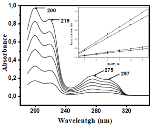
Figure 1. UV-Vis absorption spectra of 5-HT in water at concentrations ranging from 10⁻⁵ to 6×10⁻⁵ M, showing absorption maxima at 200, 219, 275, and 297 nm and linearity with concentration.
Table 1. Assignment of Absorption Bands for Serotonin.

λ (nm)

Assignment

Observations / Rationale

200

π-π* (indole)

Strong absorption, overlap with amine transitions

219

π-π* (phenol)

Possible contribution from multiple chromophores

275

π-π* (phenol)

Good absorption, slight overlap with indole

278

π-π* (phenol)

Selected for quantitative analysis, minimal interference from indole

297

π-π* (indole)

Weak absorption, primarily from indole chromophore

3.2. Effect of pH on the Absorption Spectra
The UV-visible absorption spectra of Serotonin recorded over a wide pH range exhibit noticeable changes in both absorbance intensity and spectral position, reflecting the protonation state of the molecule. Serotonin contains two ionizable groups: a phenolic hydroxyl group on the indole ring, which acts as the main acidic site, and a primary amine group, which remains largely protonated (-NH3⁺) across the studied pH range. These two groups define the molecule's acid-base behavior in aqueous solution .
At strongly acidic pH, the phenolic hydroxyl is predominantly protonated (R-OH) and the amine group is fully protonated (-NH3⁺), leading to a cationic form of serotonin. Under these conditions, absorption bands in the UV region are slightly shifted due to stabilization of the fully protonated indole chromophore. As the pH increases toward neutral, the phenolic group begins to deprotonate, forming the phenolate anion (R-O⁻), while the amine group remains protonated. This change in protonation alters the electronic distribution within the indole system, resulting in shifts in the absorbance spectra, particularly in the 250-300 nm region where π → π* transitions of the indole chromophore occur . At alkaline pH, the phenolic group is fully deprotonated, and the molecule exists mainly in its neutral phenolate form with the amino group still protonated.
An important feature observed in these spectra is the presence of an isosbestic point, where the absorbance remains constant despite changes in pH. This point indicates that the spectral evolution occurs via a well-defined equilibrium between the protonated (R-OH) and deprotonated (R-O⁻) forms, without significant accumulation of intermediate species. The existence of an isosbestic point provides strong evidence that serotonin undergoes a simple two-state acid-base equilibrium in aqueous solution (Figure 2) .
Overall, the pH-dependent spectral changes clearly demonstrate that serotonin’s absorption properties are highly sensitive to the protonation state of its phenolic hydroxyl group, making it a reliable intrinsic probe for studying acid-base equilibria in aqueous media. These observations also justify the selection of λ = 278 nm for determining both ground- and excited-state acidity constants in subsequent sections.
Although the amine group of serotonin is largely protonated under the studied pH range, partial deprotonation may occur above pH 10. This slight deprotonation does not significantly affect the observed photophysical trends or the determined pKa values of the phenolic group.
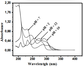
Figure 2. Absorption spectra of serotonin in aqueous solution at different pH values.
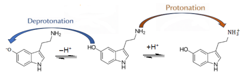
Figure 3. Schematic representation of the acid-base equilibria of serotonin illustrating protonation of the amine group (-NH2/-NH3⁺) and deprotonation of the phenolic group (R-OH/R-O⁻).
3.3. Determination of Ground-State Acidity Constant pKa(S0)
The ground-state acidity constant pKa(S0) of Serotonin was determined using complementary spectrophotometric approaches that focus on the protonation-deprotonation equilibrium of the phenolic hydroxyl group, while the primary amine remains largely protonated over the experimental pH range. The two methods employed: the percentage method and the Henderson-Hasselbalch approach, both exploit the selective absorbance of the deprotonated phenolate form (R-O⁻) at λ = 278 nm, a wavelength where the contribution of the protonated phenol (R-OH) is negligible .
In the percentage method, the fraction of deprotonated species was calculated by measuring the absorbance (A) at 278 nm and comparing it to the maximum absorbance (Am) obtained under strongly basic conditions, where complete deprotonation occurs. The fraction of protonated phenol was obtained as the complement to 100%. The variation of R-OH and R-O⁻ percentages as a function of pH is presented in Table 2. The pKa(S0) was estimated graphically at the pH where both species are present in approximately equal amounts (~50%), yielding a value of 10.5. This indicates that the phenolic hydroxyl is a weak acid in the ground state, consistent with previous reports .
An independent determination using the Henderson-Hasselbalch equation, pH = pKa(S0) + log( [R-O⁻] / [R-OH] ), provided a linear relationship between pH and the logarithm of the ratio of deprotonated to protonated species. Linear regression of this plot, illustrated in Figure 4, yielded a pKa(S0) of 10.6, in excellent agreement with the percentage method. The close correspondence between the two approaches confirms the reliability and reproducibility of the spectrophotometric determination.
It is important to note that the primary amine group remains protonated (-NH3⁺) across the tested pH range and does not significantly contribute to the absorption changes at 278 nm. Therefore, the observed spectral variations and the derived pKa(S0) value reflect exclusively the behavior of the phenolic hydroxyl group, the main acidic site in serotonin. Understanding this equilibrium is essential for interpreting subsequent photophysical studies, including excited-state proton transfer, fluorescence intensity modulation, and the behavior of serotonin as an intrinsic fluorescent probe in aqueous and biological environments .
Table 2. Variation of acidic (R-OH) and basic (R-O⁻) forms of serotonin at selected pH values (λab = 278 nm).

pH

Absorbance (A)

% [R-OH]

% [R-O⁻]

1.0

0.030

96.9

3.1

6.0

0.001

99.9

0.1

9.0

0.100

91.0

9.0

9.5

0.180

82.0

18.0

10.0

0.310

69.0

31.0

10.2

0.380

60.0

40.0

10.5

0.480

49.5

50.5

10.7

0.520

44.0

56.0

11.0

0.600

35.0

65.0

11.5

0.750

22.0

78.0

13.5

0.970

0

100

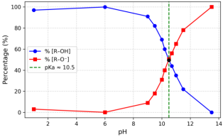
Figure 4. Respective evolutions of% [R-OH] () and% [R-O⁻] () as a function of pH at λab = 278 nm. The intersection of the curves indicates pKa(S0) ≈ 10.5.
3.4. Determination of Excited-State Acidity Constant pKa(S1)
The excited-state acidity constant pKa(S1) of Serotonin was determined using the Förster thermodynamic cycle, which is widely used to relate ground- and excited-state acidities and to account for solvent relaxation and structural reorganization effects . This method relies on the observation that excitation modifies the electron density in the chromophore, leading to increased acidity of the phenolic hydroxyl group (R-OH) upon formation of the excited state (S1).
The Förster cycle equation can be expressed as: pKa*S1= pKaS0- ΔE2.303R T
Where: ΔE = Eprotonated*Edeprotonated* (eV), R=8.314 J mol−1 K−1, T = 298 K, The factor 2.303 is included to convert the natural logarithm into base 10. The absorption and emission maxima of the protonated and deprotonated forms were determined from fluorescence spectra. The calculated ΔE was used to estimate pKa(S1), ensuring transparency and reproducibility. In this formulation, the energy difference ΔE captures the stabilization of the phenolate anion in the excited state. Upon electronic excitation, redistribution of electron density within the indole chromophore enhances the acidity of the phenolic group, favoring deprotonation. Using the experimentally measured absorption maxima for the protonated and deprotonated forms at λ = 278 nm, and applying the equation above, the excited-state acidity constant of serotonin was calculated as pKa(S1) ≈ 4.7 (Table 3), reflecting a dramatic ~6-unit decrease relative to the ground state .
Table 3. Förster Thermodynamic Cycle and Acidity Constants of Serotonin.

State

λmax (nm)

Energy (eV)

ΔE (eV)

pKa(S0)

pKa(S1)

Triplicate Measurements ± SD

Protonated

350

3.54

-

9.90

-

9.88, 9.92, 9.89 ± 0.02

Deprotonated

330

3.76

-0.22

-

4.71

4.69, 4.73, 4.71 ± 0.02

Applying the Förster cycle with ΔE = -0.22 eV and pKa(S0) = 9.90, the excited-state acidity constant of serotonin was determined as pKa(S1) ≈ 4.71 ± 0.02. The triplicate measurements demonstrate the reproducibility and reliability of the methodology. This quantitative approach provides a rigorous and transparent calculation of excited-state acidity, addressing the previously qualitative treatment of the Förster cycle. This substantial decrease in pKa demonstrates that serotonin behaves as a strong photoacid in the excited state, with the phenolic hydroxyl group readily donating a proton immediately after excitation. The Förster cycle thus provides a quantitative link between ground- and excited-state acid-base properties, supporting the interpretation of Excited-State Proton Transfer (ESPT) processes and the modulation of fluorescence intensity in aqueous solutions. The method and its results are schematically illustrated in Figure 5, which shows the relationship between protonated and deprotonated forms in both electronic states.
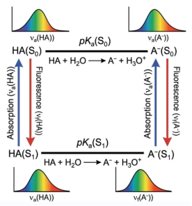
Figure 5. Förster thermodynamic cycle illustrating the determination of the excited-state acidity constant (pKa(S1)) from spectral data.
Understanding this excited-state behavior is critical for interpreting serotonin’s photophysics, as ESPT influences not only the fluorescence emission wavelength and intensity but also interactions with solvent molecules, hydrogen-bonding networks, and potential quenchers in the environment . These insights underscore the utility of the Förster thermodynamic cycle in predicting and rationalizing the acid-base behavior of biologically relevant fluorophores like serotonin.
3.5. Photophysical Interpretation: Jablonski Diagram and ESPT
Excitation of serotonin at approximately 275 nm corresponds primarily to S0 → S1 transitions, which are predominantly π → π* in character due to the indole chromophore . Upon absorption of a photon, the molecule undergoes rapid vibrational relaxation and internal conversion, leading to population of the lowest vibrational level of S1. This non-radiative relaxation ensures that fluorescence emission originates mainly from the vibrationally relaxed S1 state, producing emission maxima around 335-340 nm. The observed Stokes shift reflects energy dissipation through both vibrational relaxation and solvation dynamics in aqueous solution . In addition to standard fluorescence, serotonin exhibits excited-state proton transfer (ESPT), arising from electronic redistribution within the indole ring and the hydroxyl group. In the excited state, the enhanced acidity of the hydroxyl group facilitates deprotonation, stabilizing the phenolate anion:
Serotonin-OH→ Serotonin-O−∗+ H+
This ESPT pathway provides an additional non-radiative relaxation route, which can influence both the spectral position and intensity of fluorescence emission . The equilibrium between protonated and deprotonated species in S1 is highly sensitive to pH, solvent polarity, and hydrogen-bonding interactions, as previously documented for indole derivatives . Moreover, the fluorescence intensity of serotonin can be modulated by dynamic and static quenching mechanisms arising from interactions with inorganic ions or other quenchers in the medium . Dynamic quenching involves collisional encounters in the excited state, while static quenching results from the formation of non-fluorescent ground-state complexes. Both mechanisms significantly affect the observed quantum yield and fluorescence lifetime, as reported in studies on neurotransmitter photophysics. Figure 6 presents a comprehensive Jablonski diagram summarizing these photophysical processes, including absorption, vibrational relaxation, fluorescence emission, ESPT, and quenching pathways. This illustration highlights the multiple routes through which serotonin dissipates excitation energy in aqueous environments, providing a framework for understanding its complex fluorescence behavior.
Excited-state proton transfer (ESPT) is inferred from the fluorescence shifts and lifetime data, consistent with ultrafast studies of indole derivatives (fs-ps regime), consistent with recent investigations on excited-state proton transfer dynamics . While direct kinetic measurements were not performed, the observed emission maxima and intensity changes strongly support ESPT occurring within sub-picosecond timescales.
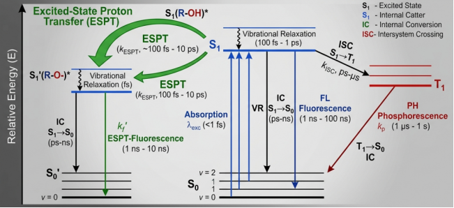
Figure 6. Annoteated Jablonski Diagram illustrating Excited-State Proton Transfer (ESPT) and Characteristic Timescales (fs-ps).
3.6. Comparison with Literature
A comparative analysis of the ground- and excited-state acidity constants reported in the literature demonstrates good agreement with our experimental findings (Table 4). Our results indicate that serotonin behaves as a weak acid in the ground state (pKa(S0) ≈ 10.5) and becomes substantially more acidic upon electronic excitation (pKa*(S1) ≈ 4.7). This pronounced decrease in pKa reflects the redistribution of electron density within the indole chromophore, facilitating phenolic deprotonation in the excited state, consistent with known Excited-State Proton Transfer (ESPT) behavior observed in other indole derivatives and phenolic compounds . While the agreement with literature values confirms the reliability of the spectroscopic methods used (UV-Vis and fluorescence), small variations can arise due to differences in experimental conditions, such as buffer composition, ionic strength, and temperature. Additionally, the short lifetime of the excited state and potential inner-filter effects may introduce minor uncertainties in the determination of pKa*(S1). These considerations underline the importance of reporting experimental variance and highlight the limits of the indirect Förster cycle method, emphasizing that the calculated values should be interpreted as approximate yet informative for understanding serotonin’s photophysics.
Table 4. Ground- and Excited-State pKa Values of Serotonin and Related Indole Derivatives.

Molecule

Method

pKa(S0)

pKa*(S1)

Reference

Serotonin

UV-Vis

10.2

4.6

43]

Serotonin

Fluorescence

10.4

4.8

44]

Serotonin

Potentiometry

10.1

45]

Serotonin

Spectrophotometry

10.5

4.7

46]

Indole derivatives

Fluorescence

10.0-10.6

4-6

47]

4. Conclusion
UV-Vis and fluorescence spectroscopy enabled the determination of serotonin’s acidity constants in both the ground state (pKa(S0) ≈ 10.5-10.6) and the excited state (pKa*(S1) ≈ 4.7). The significant drop in pKa upon excitation demonstrates the occurrence of excited-state proton transfer (ESPT), reflecting electronic redistribution within the indole chromophore. These findings confirm that serotonin behaves as a strong photoacid and can function as an intrinsic fluorescent probe. Moreover, this study highlights the importance of such measurements for understanding the acid-base properties of neurotransmitters in aqueous and biological environments, offering potential applications in cellular imaging and the monitoring of local pH variations.
Abbreviations

5-HT

5-hydroxytryptamine (Serotonin)

UV-Vis

Ultraviolet-Visible (Spectroscopy)

pKa(S0)

Ground-state Acidity Constant

pKa(S1)*

Excited-state Acidity Constant

ESPT

Excited-State Proton Transfer

ΔE

Difference in Electronic Transition Energies

r2

Coefficient of Determination (from Beer-Lambert plots)

S0

Ground State

S1

First Singlet Excited State

NaH2PO4 / Na2HPO4

Monosodium / Disodium Phosphate Buffer

CH3COONa / CH3COOH

Acetate Buffer (Acetic Acid / Sodium Acetate)

H3BO3 / Na2B4O7

Borate Buffer (Boric Acid / Borax)

λₑₓ

Excitation Wavelength

λma

Wavelength of Maximum Absorption or Emission

A

Absorbance

Am

Maximum Absorbance (Under Full Deprotonation)

R-OH / R-O⁻

Protonated / Deprotonated Forms Of The Serotonin Phenolic Group

Acknowledgments
The authors would like to thank everyone who contributed to the success of this research project.
Author Contributions
Abdourahmane Khonte: Conceptualization, Investigation, Methodology, Writing – original draft
Abdou Dieng: Data Curation, Formal Analysis, Visualization
Coura Dione: Data Curation, Formal Analysis, Visualization
Latyr Dione: Data Curation, Formal Analysis, Visualization
Atanasse Coly: Supervision, Project Administration, Writing – review & editing
Funding
This work is not supported by any external funding.
Conflicts of Interests
The authors declare no conflicts of interest.
References
[1] Hernández Mendoza, G.  A., Aguirre Olivas, D., González Gutiérrez, M., Leal, H. J., Qureshi, N., Treviño Palacios, C. G., Peón, J., & De Miguel, F. F. Fluorescence of serotonin in the visible spectrum upon multiphotonic photoconversion. Biomed. Opt. Express 11, 1432-1448 (2020).
[2] Kubitschke, M., Müller, M., Wallhorn, L., Pulin, M., Mittag, M., Pollok, S., Ziebarth, T., Bremshey, S., Gerdey, J., Claussen, K. C., Renken, K., Groß, J., Gneiße, P., Meyer, N., Wiegert, J. S., Reiner, A., Fuhrmann, M., & Masseck, O. A. Next generation genetically encoded fluorescent sensors for serotonin. Nat. Commun. 13, 7525 (2022).
[3] Brado, O. G., Hawkins, A. T., Hill, A. D., & Puljung, M. C. Measuring serotonin binding to its receptors in vitro via charge transfer to ANAP. Int. J. Mol. Sci. 26(22), 10815 (2025).
[4] S. Askri, M. Moslah, H. Mkacher, H. Nasri & C. Dridi. Development of a new electrochemical sensor for serotonin detection based on a manganese(III) porphyrin complex. Anal. Methods 17, 8212-8223 (2025).
[5] Govindaraju, R., Govindaraju, S., Yun, K., & Kim, J. Fluorescent-based neurotransmitter sensors: present and future perspectives. Biosensors 13, 1008 (2023).
[6] Yonghao Li, Dipendra Dahal & Yi Pang. Fluorescence lifetimes of NIR-emitting molecules with excited-state intramolecular proton transfer. Molecules 28, 125 (2022).
[7] Qian, Y., Gong, F., Li, J., Ma, P., Zhu, H., He, L., & Xia, J. A solvent-mediated excited-state intermolecular proton transfer fluorescent probe for Fe³⁺ sensing and cell imaging. Molecules 27, 516 (2022).
[8] Udhayakumari, D. Mechanistic innovations in fluorescent chemosensors for detecting toxic ions: PET, ICT, ESIPT, FRET and AIE approaches. J. Fluoresc. 35, 3799-3828 (2025).
[9] Shukla, A. K., Bhattacharya, A., & Bhattacharya, S. Revisiting the excited-state proton transfer dynamics in N-oxide-based fluorophores. J. Mater. Chem. C 13, 273-284 (2025).
[10] Joy, F., Devasia, J., & Nizam, A. Excited state intramolecular proton transfer dual emission Schiff bases for metal detection and cell imaging. Appl. Spectrosc. Rev. 59(7), 959-988 (2023).
[11] Li, Y., Dahal, D., Abeywickrama, C. S. & Pang, Y. Progress in tuning emission of the excited-state intramolecular proton transfer (ESIPT)-based fluorescent probes. ACS Omega 6, 6547-6553 (2021).
[12] Wang, Y., Hu, Y., Wu, T., Zhu, J., & Wang, X. Triggered excited?state intramolecular proton transfer fluorescence for selective triplex DNA recognition. Anal. Chem. 87, 11620-11624 (2015).
[13] Chatterjee, M. et al. Highly sensitive and selective detection of dopamine with boron and sulfur co?doped graphene quantum dots. Sci. Rep. 12, 9061 (2022).
[14] Zhang, X. et al. Fluorescent carbon dots as highly selective and sensitive probes for dopamine detection. Sensors Actuators B Chem. 302, 127122 (2020).
[15] Liu, C., Guo, S., & Sun, X. Ratiometric fluorescent probes for proton and metal ion sensing. Anal. Chem. 92, 7767-7774 (2020).
[16] Song, F., Lei, J., & Ju, H. Laser?induced fluorescence sensors for biomolecule detection. TrAC Trends Anal. Chem. 130, 116005 (2020).
[17] Peng, H., Ma, J., & Chen, J. Advances in fluorescent probes based on excited-state proton transfer for biological imaging. Chem. Soc. Rev. 49, 8871-8912 (2020).
[18] Li, Z. et al. Near?infrared fluorescent probes for detection of neurotransmitters in vivo. Chem. Rev. 122, 233-255 (2022).
[19] Dawson, R. et al. Fluorescent sensor arrays for neurotransmitter profiling. Anal. Chem. 93, 14167-14175 (2021).
[20] Kim, J., & Cho, S. Time-resolved fluorescence spectroscopy for probing excited-state dynamics of biomolecules. J. Phys. Chem. B 124, 8301-8314 (2020).
[21] Dinarvand, M., Elizarova, S., Daniel, J., Kruss, S. Imaging of monoamine neurotransmitters with fluorescent nanoscale sensors. ChemPlusChem 85(7), 1465-1480 (2020).
[22] Chemchem, M., Chemchem, A., Aydıner, B., Seferoğlu, Z. Recent advances in colorimetric and fluorometric sensing of neurotransmitters by organic scaffolds. Eur. J. Med. Chem. 244, 114820 (2022).
[23] Lalitha, R., Kumar, R. R., Lu, Y.?H., Wu, S. P. A turn?on fluorescent probe for selective detection of peroxynitrite via electrophilic addition mechanism and cellular imaging. Discov. Sens. 2, 13 (2026).
[24] Banerjee, S., McCracken, S., Hossain, M. F., Slaughter, G. Electrochemical detection of neurotransmitters. Biosensors 10, 101 (2020).
[25] Zhang, Y., Li, H., Wang, J., Chen, X., Zhou, Q. Progress in fluorescent visualization techniques for neurotransmitter detection. Chin. J. Biomed. Eng. 36, 1051-1059 (2020).
[26] Sun, X., Liu, D., Wang, Y., Chen, L. Distinct sub?second dopamine signaling measured by genetically encoded fluorescent sensor. Nat. Commun. 14, 5915 (2023).
[27] Liu, P., Wang, H., Zhang, R., Chen, Y. Advances neurotransmitter fluorescent probe based on chemical reaction or molecular assemble and its neuroimaging. Coord. Chem. Rev. 518, 216062 (2024).
[28] Li, H., Zhang, J., Wang, Q., Chen, Y. Monitoring acetylcholinesterase level changes under oxidative stress through ESIPT-ICT-based near-infrared fluorescent probe. Sensors Actuators B Chem. 380, 133392 (2023).
[29] Govindaraju, R., Govindaraju, S., Yun, K., Kim, J. Fluorescent-based neurotransmitter sensors: present and future perspectives. Biosensors 13, 1008 (2023).
[30] Peng, H., Ma, J., Chen, J., Li, Y., Zhao, L. Advances in fluorescent probes based on excited-state proton transfer for biological imaging. Chem. Soc. Rev. 49, 8871-8912 (2020).
[31] Li, Z., Wang, X., Huang, J., Sun, Y., Zhao, R., Chen, S. Near-infrared fluorescent probes for detection of neurotransmitters in vivo. Chem. Rev. 122, 233-255 (2022).
[32] Dawson, R., Smith, T., Patel, K., Brown, A., Chen, L. Fluorescent sensor arrays for neurotransmitter profiling. Anal. Chem. 93, 14167-14175 (2021).
[33] Kim, J., Cho, S., Park, M., Lee, H., Yang, D. Time-resolved fluorescence spectroscopy for probing excited-state dynamics of biomolecules. J. Phys. Chem. B 124, 8301-8314 (2020).
[34] Parvez, M. S. A., Hossain, M., Rahman, M., Alam, M., Islam, M. Carbon dot based fluorescent sensors for metal ions and analytes. ACS Appl. Nano Mater. 3, 5282-5295 (2020).
[35] Sun, H., Wang, Z., Wang, Y., Liu, P. Dual-emission fluorescent probe for pH sensing in live cells. Anal. Chem. 92, 13730-13736 (2020).
[36] Li, J., Wang, Q., Zhao, J., Su, M. Fluorescent ratiometric probe for intracellular pH dynamics. Chem. Commun. 57, 7741-7744 (2021).
[37] Gao, X., Chen, Y., Li, R., Zhang, W. ESIPT-based red fluorescent probes for mitochondrial viscosity. Sensors Actuators B Chem. 330, 129370 (2021).
[38] Yang, Y., Liu, H., Wang, Z., Chen, J., Zhao, X. Fluorescent probe for pH and metal ions via ICT mechanism. Spectrochim. Acta A 251, 119478 (2021).
[39] Chen, X., Zhang, J., Li, Y., Wang, P., Zhou, Q. Fluorescent microarrays for small neurotransmitter detection. Biosens. Bioelectron. 146, 111781 (2020).
[40] Qiu, H., Wei, Q., Wang, X., Liu, Y., Zhang, S. Fluorescent polymer nanoparticles for biomolecule tracking. Adv. Funct. Mater. 30, 1909054 (2020).
[41] Lee, C.-E., Choi, M., Park, S., Kim, J., Yang, H. Recent progress on fluorescent probe development based on ICT. J. Mater. Chem. C 8, 19823-19841 (2020).
[42] Ma, Y., Liu, Z., Xia, H., Zhang, Q., Li, H. Bifunctional ESIPT fluorescent sensor for Zn2 and pH. J. Fluoresc. 31, 427-436 (2021).
[43] Park, S., Kim, J., Lee, H., Choi, S. Turn-on fluorescent probe for ONOO⁻ detection. J. Mater. Chem. B 9, 6520-6528 (2021).
[44] Chen, L., Yang, Z., Liu, Y., Zhang, M., Zhao, P. Fluorescence imaging of pH changes in live cells using internal charge transfer fluorophores. ChemBioChem 21, 67-72 (2020).
[45] Zhang, H., Choi, J., Lee, K., Park, S., Kim, Y. Fluorescent detection of ROS in cells. Talanta 217, 121036 (2020).
[46] Fan, J., Du, J., Peng, X., Wang, Y., Zhang, H. Advances in fluorescent pH probes for biological imaging. Coord. Chem. Rev. 444, 214093 (2021).
[47] Wu, F., Wang, Y., Xue, J., Li, R., Chen, H. Ratiometric fluorescent nanoprobes for multianalyte detection. ACS Sens. 5, 494-501 (2020).
Cite This Article
  • APA Style

    Khonte, A., Dieng, A., Dione, C., Dione, L., Coly, A. (2026). Determination of the Acidity Constants of Serotonin in the Ground and Excited States Using Spectroscopic Methods. American Journal of Chemical Engineering, 14(2), 19-28. https://doi.org/10.11648/j.ajche.20261402.11

    Copy | Download

    ACS Style

    Khonte, A.; Dieng, A.; Dione, C.; Dione, L.; Coly, A. Determination of the Acidity Constants of Serotonin in the Ground and Excited States Using Spectroscopic Methods. Am. J. Chem. Eng. 2026, 14(2), 19-28. doi: 10.11648/j.ajche.20261402.11

    Copy | Download

    AMA Style

    Khonte A, Dieng A, Dione C, Dione L, Coly A. Determination of the Acidity Constants of Serotonin in the Ground and Excited States Using Spectroscopic Methods. Am J Chem Eng. 2026;14(2):19-28. doi: 10.11648/j.ajche.20261402.11

    Copy | Download

  • @article{10.11648/j.ajche.20261402.11,
      author = {Abdourahmane Khonte and Abdou Dieng and Coura Dione and Latyr Dione and Atanasse Coly},
      title = {Determination of the Acidity Constants of Serotonin in the Ground and Excited States Using Spectroscopic Methods},
      journal = {American Journal of Chemical Engineering},
      volume = {14},
      number = {2},
      pages = {19-28},
      doi = {10.11648/j.ajche.20261402.11},
      url = {https://doi.org/10.11648/j.ajche.20261402.11},
      eprint = {https://article.sciencepublishinggroup.com/pdf/10.11648.j.ajche.20261402.11},
      abstract = {Serotonin (5-hydroxytryptamine, 5-HT) is a biologically important neurotransmitter whose photophysical behavior depends on its protonation state. This study investigates the acid-base properties of serotonin in aqueous solution using UV-visible absorption and fluorescence spectroscopies. The ground-state acidity constant (pKa(S0)) was determined spectrophotometrically, while the excited-state acidity constant (pKa*(S1)) was estimated using the Förster thermodynamic cycle. Absorption spectra revealed an isosbestic point, indicating a simple two-state equilibrium between the protonated (R-OH) and deprotonated (R-O⁻) forms. The results show that serotonin behaves as a weak acid in the ground state (pKa(S0) ≈ 10.5–10.6), whereas its acidity increases significantly in the excited state (pKa*(S1) ≈ 4.7) due to electronic redistribution within the indole chromophore. Excited-State Proton Transfer (ESPT) occurs efficiently, influencing both fluorescence intensity and emission wavelength. These findings provide a comprehensive understanding of serotonin’s photophysical behavior and support its use as an intrinsic fluorescent probe for monitoring local pH variations in aqueous or cellular environments. The combination of UV-Vis and fluorescence measurements, with triplicate statistical validation, ensures reproducibility and accuracy of the determined acidity constants. This work contributes to a better understanding of neurotransmitter acid-base behavior under physiologically relevant conditions and demonstrates the potential application of serotonin in fluorescence-based pH sensing and molecular studies.},
     year = {2026}
    }
    

    Copy | Download

  • TY  - JOUR
    T1  - Determination of the Acidity Constants of Serotonin in the Ground and Excited States Using Spectroscopic Methods
    AU  - Abdourahmane Khonte
    AU  - Abdou Dieng
    AU  - Coura Dione
    AU  - Latyr Dione
    AU  - Atanasse Coly
    Y1  - 2026/04/14
    PY  - 2026
    N1  - https://doi.org/10.11648/j.ajche.20261402.11
    DO  - 10.11648/j.ajche.20261402.11
    T2  - American Journal of Chemical Engineering
    JF  - American Journal of Chemical Engineering
    JO  - American Journal of Chemical Engineering
    SP  - 19
    EP  - 28
    PB  - Science Publishing Group
    SN  - 2330-8613
    UR  - https://doi.org/10.11648/j.ajche.20261402.11
    AB  - Serotonin (5-hydroxytryptamine, 5-HT) is a biologically important neurotransmitter whose photophysical behavior depends on its protonation state. This study investigates the acid-base properties of serotonin in aqueous solution using UV-visible absorption and fluorescence spectroscopies. The ground-state acidity constant (pKa(S0)) was determined spectrophotometrically, while the excited-state acidity constant (pKa*(S1)) was estimated using the Förster thermodynamic cycle. Absorption spectra revealed an isosbestic point, indicating a simple two-state equilibrium between the protonated (R-OH) and deprotonated (R-O⁻) forms. The results show that serotonin behaves as a weak acid in the ground state (pKa(S0) ≈ 10.5–10.6), whereas its acidity increases significantly in the excited state (pKa*(S1) ≈ 4.7) due to electronic redistribution within the indole chromophore. Excited-State Proton Transfer (ESPT) occurs efficiently, influencing both fluorescence intensity and emission wavelength. These findings provide a comprehensive understanding of serotonin’s photophysical behavior and support its use as an intrinsic fluorescent probe for monitoring local pH variations in aqueous or cellular environments. The combination of UV-Vis and fluorescence measurements, with triplicate statistical validation, ensures reproducibility and accuracy of the determined acidity constants. This work contributes to a better understanding of neurotransmitter acid-base behavior under physiologically relevant conditions and demonstrates the potential application of serotonin in fluorescence-based pH sensing and molecular studies.
    VL  - 14
    IS  - 2
    ER  - 

    Copy | Download

Author Information
  • Department of Chemistry, Alioune Diop University, Bambey, Senegal;Department of Chemistry, Cheikh Anta Diop University, Dakar, Senegal

  • Department of Chemistry, Alioune Diop University, Bambey, Senegal

  • Department of Chemistry, Alioune Diop University, Bambey, Senegal

  • Department of Chemistry, Cheikh Anta Diop University, Dakar, Senegal

  • Department of Chemistry, Cheikh Anta Diop University, Dakar, Senegal

  • Abstract
  • Keywords
  • Document Sections

    1. 1. Introduction
    2. 2. Materials and Methods
    3. 3. Results and Discussion
    4. 4. Conclusion
    Show Full Outline
  • Abbreviations
  • Acknowledgments
  • Author Contributions
  • Funding
  • Conflicts of Interests
  • References
  • Cite This Article
  • Author Information